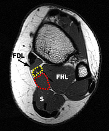
Foot Muscles Mri - Accessory Muscles Anatomy Symptoms And Radiologic Evaluation Radiographics. The majority of soft tissue lesions in the foot and ankle are benign. The three plantar interossei muscles adduct the 3 rd, 4 th and 5 th toes toward the long axis through the 2 nd toe. Trauma effects of direct injury or tear denervation injury: Mri of the ankle and feet Proper interpretation of the findings is crucial, especially in elite athletes.
An ankle mri also offers a look at the bones of the lower leg that help make up the ankle joint, such as the tibia and fibula, as well as the muscles of the foot. Anatomical structures of the ankle and foot and specific regions (major joints) are visible as dynamic labeled images. Findings on conventional arthrography and mr imaging. • muscle edema is seen secondary to multiple etiologies including trauma, infectious and inflammatory processes, autoimmune disorders, neoplasms, and denervation injuries • on mri muscle edema is characterized by increase in free water within the muscle • muscle edema is seen on mri as increased signal on fluid sensitive sequences t2 fs They are named extensor digitorum brevis and extensor hallucis brevis.
Http Pdf Posterng Netkey At Download Index Php Module Get Pdf By Id Poster Id 40701 from Chronic plantar fasciitis may be accompanied by muscle atrophy of plantar intrinsic foot muscles and tibialis posterior compromising the dynamic support of the foot prolonging the injury. Like the fingers, the toes have flexor and extensor muscles that power their movement and play a large. Muscle injuries of the hip and thigh are a highly relevant issue in competitive sports imaging. An extremity mri is a type of scan used specifically for diagnostic imaging of the arm, leg, hand, or foot. Mri findings of acute turf toe: Magnetic resonance imaging (mri) is the modality of choice in diagnosing accessory muscles, delineating their relationship to adjacent structures, and differentiating them from soft tissue tumors. The machine uses radio waves and a magnetic field to generate images of the inside of the extremity in order to diagnose problems with the muscles, bones, joints, nerves, or blood vessels. Routine ankle magnetic resonance imaging (mri) tests involve taking images of the foot and ankle in the axial, coronal, and sagittal planes parallel to the tabletop(2).
Anatomical structures of the ankle and foot and specific regions (major joints) are visible as dynamic labeled images.
Mri is the choice of modality for further imaging the ankle and foot after obtaining initial radiographs. Radiologists need to be familiar with typical mri findings in order to accurately detect and classify muscle injuries. Muscle injuries of the hip and thigh are a highly relevant issue in competitive sports imaging. A case report and review of anatomy. Like the fingers, the toes have flexor and extensor muscles that power their movement and play a large. The majority of soft tissue lesions in the foot and ankle are benign. They are mainly responsible for assisting some of the extrinsic muscles in their actions. 9 yao l, do hm, cracchiolo a, et al. Magnetic resonance imaging (mri) is the modality of choice in diagnosing accessory muscles, delineating their relationship to adjacent structures, and differentiating them from soft tissue tumors. They are named extensor digitorum brevis and extensor hallucis brevis. Six hundred two patients were included in the study: • muscle edema is seen secondary to multiple etiologies including trauma, infectious and inflammatory processes, autoimmune disorders, neoplasms, and denervation injuries • on mri muscle edema is characterized by increase in free water within the muscle • muscle edema is seen on mri as increased signal on fluid sensitive sequences t2 fs 23,25 mri at the level of the malleolus demonstrates the muscle as.
They are mainly responsible for assisting some of the extrinsic muscles in their actions. The most common ossicle is the os trigonum, which is a prominent unfused apophysis of the lateral tubercle of the talus. The aim of this review is to provide the reader with a comprehensive overview of the magnetic resonance imaging (mri) characteristics of the most common benign and malignant soft tissue neoplasms which occur around the foot and ankle. The machine uses radio waves and a magnetic field to generate images of the inside of the extremity in order to diagnose problems with the muscles, bones, joints, nerves, or blood vessels. Your doctor, with the help of a radiologist, can then examine these images to determine whether there is anything wrong with your foot or ankle.
Accessory Muscles Of The Ankle Radsource from radsource.us With a muscle injury, for example, mri images often show a bright signal indicating that there is more water in the muscle, which is a sign of injury. The majority of soft tissue lesions in the foot and ankle are benign. Magnetic resonance imaging, otherwise known as mri, uses a combination of magnetic fields and radio waves to take images of the internal structures of your body. The muscles lie within a flat fascia on the dorsum of the foot (fascia dorsalis pedis) and are innervated by the deep fibular or peroneal nerve. All images were evaluated for the presence of selective fatty atrophy of the adq muscle. They are named extensor digitorum brevis and extensor hallucis brevis. A prospective study of all patients referred for ankle and foot mri examinations was performed. This imaging technique assesses the ligaments and tendons, neurovascular structures (tarsal tunnel and plantar fascia), and the osseous structures(19).
• muscle edema is seen secondary to multiple etiologies including trauma, infectious and inflammatory processes, autoimmune disorders, neoplasms, and denervation injuries • on mri muscle edema is characterized by increase in free water within the muscle • muscle edema is seen on mri as increased signal on fluid sensitive sequences t2 fs
Anatomical structures of the ankle and foot and specific regions (major joints) are visible as dynamic labeled images. This imaging technique assesses the ligaments and tendons, neurovascular structures (tarsal tunnel and plantar fascia), and the osseous structures(19). Magnetic resonance imaging, otherwise known as mri, uses a combination of magnetic fields and radio waves to take images of the internal structures of your body. Routine ankle magnetic resonance imaging (mri) tests involve taking images of the foot and ankle in the axial, coronal, and sagittal planes parallel to the tabletop(2). The peroneus quartus muscle is more common, presenting in 13% to 22% of the population. The presence of intramuscular edema (increased high t2/stir signal) on mri carries an extremely broad differential. The most common ossicle is the os trigonum, which is a prominent unfused apophysis of the lateral tubercle of the talus. Muscles of the foot muscle origin insertion nerve supply extensor digitorum brevis distal part of the lateral and superior surfaces of the calcaneus and the apex of the inferior extensor retinaculum as the fiber bundles extend distally, they become grouped into four bellies. In the foot and ankle many accessory ossicles can be seen. Your doctor, with the help of a radiologist, can then examine these images to determine whether there is anything wrong with your foot or ankle. The aim of this review is to provide the reader with a comprehensive overview of the magnetic resonance imaging (mri) characteristics of the most common benign and malignant soft tissue neoplasms which occur around the foot and ankle. A case report and review of anatomy. The majority of soft tissue lesions in the foot and ankle are benign.
The machine uses radio waves and a magnetic field to generate images of the inside of the extremity in order to diagnose problems with the muscles, bones, joints, nerves, or blood vessels. A prospective study of all patients referred for ankle and foot mri examinations was performed. Trauma effects of direct injury or tear denervation injury: The most common ossicle is the os trigonum, which is a prominent unfused apophysis of the lateral tubercle of the talus. In the foot and ankle many accessory ossicles can be seen.
Soft Tissues May Help Determine Response To Ponseti Treatment Lerpediatrics Com from lermagazine.com The machine uses radio waves and a magnetic field to generate images of the inside of the extremity in order to diagnose problems with the muscles, bones, joints, nerves, or blood vessels. 9 yao l, do hm, cracchiolo a, et al. Related posts of foot muscle anatomy mri muscle anatomy trivia. The muscles lie within a flat fascia on the dorsum of the foot (fascia dorsalis pedis) and are innervated by the deep fibular or peroneal nerve. Both muscles are innervated by the deep fibular nerve. The peroneus quartus muscle is more common, presenting in 13% to 22% of the population. Adductor hallucis is anatomically located in the central compartment of foot, but the muscle is functionally grouped with the medial plantar muscles of foot because it acts on the great toe (hallux). • muscle edema is seen secondary to multiple etiologies including trauma, infectious and inflammatory processes, autoimmune disorders, neoplasms, and denervation injuries • on mri muscle edema is characterized by increase in free water within the muscle • muscle edema is seen on mri as increased signal on fluid sensitive sequences t2 fs
The aim of this review is to provide the reader with a comprehensive overview of the magnetic resonance imaging (mri) characteristics of the most common benign and malignant soft tissue neoplasms which occur around the foot and ankle.
9 yao l, do hm, cracchiolo a, et al. The presence of intramuscular edema (increased high t2/stir signal) on mri carries an extremely broad differential. In the foot and ankle many accessory ossicles can be seen. All images were evaluated for the presence of selective fatty atrophy of the adq muscle. Those fibers of the most medial and largest belly are… Mri is the choice of modality for further imaging the ankle and foot after obtaining initial radiographs. These findings are important and relevant for clinicians involved in the assessment and treatment of foot and lower. Mri is an ideal method for identifying areas of muscle atrophy and fatty infiltration. In addition, an image of all the muscles of the back and plantar part of the foot, all tendons and tendon ligaments, blood vessels and nerves are obtained. Your doctor, with the help of a radiologist, can then examine these images to determine whether there is anything wrong with your foot or ankle. The majority of soft tissue lesions in the foot and ankle are benign. Fractures or breaks in the lower portion of the tibia and fibula will show up. The machine uses radio waves and a magnetic field to generate images of the inside of the extremity in order to diagnose problems with the muscles, bones, joints, nerves, or blood vessels.